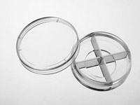
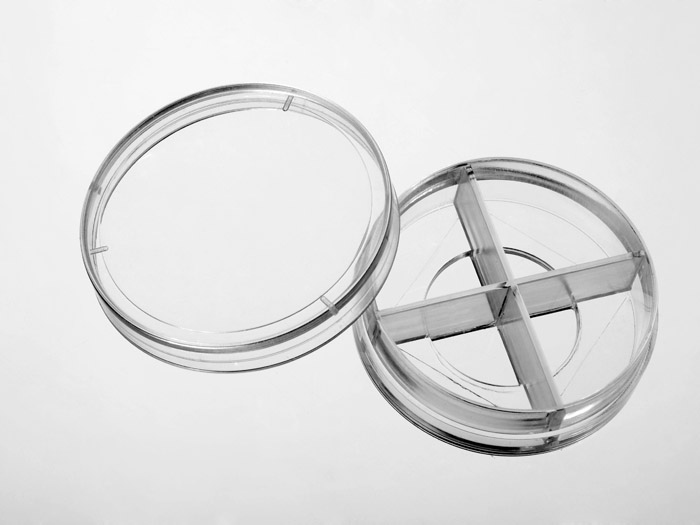
4-Chamber 35mm glass bottom dish with 20 mm microwell, #1 cover glass


35 mm glass bottom dish with 4 chambers, 20mm microwell, #1 cover glass (0.13-0.16mm). Designed for high resolution imaging such as confocal microscopy.
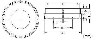
Note: We found that a small percentage of microscope adapters are too small for our 35 mm glass bottom dishes. Please check carefully the dimension diagram. If your adapter is too small, you should use our 29 mm glass bottom dish instead.
15 cases in stock
Features:
- Suitable for long term tissue culture
- Manufactured in a class 100,000 clean room
- Dish made from virgin polystyrene, tissue culture treated.
- German cover glass of superior optical quality
- A USP class VI adhesive is used to assemble the cover glass and the dish.
- Packed in easy to open peelable bag
- Sterilized by Gamma radiation.
Suitable for:
- Differential Interference Contrast (DIC)
- Widefield Fluorescence
- Confocal Microscopy
- Two-Photon and Multiphoton Microscopy
- Fluorescence Recovery After Photobleaching (FRAP)
- Förster Resonance Energy Transfer (FRET)
- Fluorescence Lifetime Imaging Microscopy (FLIM)
- Total Internal Reflection Fluorescence (TIRF)
- Super-Resolution Microscopy
Technical specifications
» View technical specification of different coverslips.
| Coverslip | #1 cover glass (0.13-0.16 mm) |
|---|---|
| Temperature Range | -20°C to 50°C |
Dimension diagram (units in mm)

Cited Publications before 2019 (3)
-
Foxo1 nucleo-cytoplasmic distribution and unidirectional nuclear influx are the same in nuclei in a single skeletal muscle fiber but vary between fibers
Y Liu, et al., American Journal of Physiology, Nov 2017
Quote: "Fibers 143 were gently dissociated and individual muscle fibers (~40/chamber) were plated on a four 144 chamber glass bottom dish (In Vitro Scientific D35C4-20-1-N) with laminin coating (Invitrogen 145 23017-015)." -
Minus end-directed kinesin-14 KIFC1 regulates the positioning and architecture of the Golgi apparatus
ZY She, et al., Oncotarget. 2017 May 30; 8(22): 36469–36483.
Quote: "Time-lapse microscopy and fluorescence recovery after photo-bleaching. Cells were grown on 12×12 mm glass coverslips in 35mm plate (glass bottom; Cellvis; Cat no. D35C4-20-1-N)" -
Tumor-stromal cross-talk: Direct cell-to-cell transfer of oncogenic microRNAs via tunneling nanotubes
V Thayanithy, et al., The Journal of Laboratory and Clinical Research, May 2014
Quote: "miR-19a transfected K7M2 cells (4 x10 5 cells) were plated on to the 4 chambered 35mm petri dishes with glass bottom (InvitroSci, Sunnyvale, CA; Catl no: D35C4-20-1-N) and allowed to adhere at standard cell culture conditions for 16 hrs and imaged by live cell microscopy"


