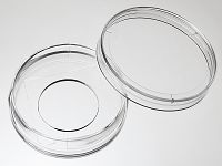
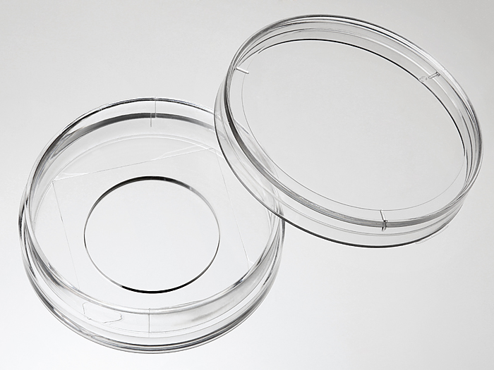
35 mm Glass bottom dish with 20 mm micro-well #1 cover glass


35mm glass bottom dish, dish size 35mm, well size 20mm, #1 cover glass(0.13mm-0.16mm). Designed for high resolution imaging such as confocal microscopy.
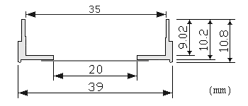
Note: We found that a small percentage of microscope adapters are too small for our 35 mm glass bottom dishes. Please check carefully the dimension diagram. If your adapter is too small, you should use our 29 mm glass bottom dish instead.
21 cases in stock
Features:
- Suitable for long term tissue culture
- Manufactured in a class 100,000 clean room
- Dish made from virgin polystyrene, tissue culture treated.
- German cover glass of superior optical quality
- A USP class VI adhesive is used to assemble the cover glass and the dish.
- Packed in easy to open peelable bag
- Sterilized by Gamma radiation.
Suitable for:
- Differential Interference Contrast (DIC)
- Widefield Fluorescence
- Confocal Microscopy
- Two-Photon and Multiphoton Microscopy
- Fluorescence Recovery After Photobleaching (FRAP)
- Förster Resonance Energy Transfer (FRET)
- Fluorescence Lifetime Imaging Microscopy (FLIM)
- Total Internal Reflection Fluorescence (TIRF)
- Super-Resolution Microscopy
Technical specifications
» View technical specification of different coverslips.
| Coverslip | #1 cover glass (0.13 - 0.16 mm) |
|---|---|
| Temperature Range | -20°C to 50°C |
| Lid diameter(outer) | 40 mm |
Dimension diagram (units in mm)

Cited Publications before 2019 (15)
-
Frequency-encoded multicolor fluorescence imaging with single-photon-counting color-blind detection
ET Garbacik, et al., Biophysical Journal, Volume 115, Issue 4, 21 August 2018, Pages 725-736
Quote: "HeLa-MannII-GFP cells were seeded on a 35-mm glass-bottomed dish with 20-mm microwell #1 cover (Cellvis, Mountain View, CA) 24 h before fixation" -
Tumor-targeting CuS nanoparticles for multimodal imaging and guided photothermal therapy of lymph node metastasis
H Shi, et al., Acta Biomaterialia Volume 72, May 2018, Pages 256-265
Quote: "MKN45 Cells (∼5 × 10 4 ) were then plated onto a glass bottom dish (In Vitro Scientific, D35-20–1-N), and allowed growing overnight." -
Microstructural properties of potato chips
S Dhital, et al., Food Structure Volume 16, April 2018, Pages 17-26
Quote: "A piece of FITC labelled and washed PC and FPC sample was placed on the 35 mm glass bottom dishes (D35-20-1-N, Cellvis, Mountain View, CA) and inundated with 50 ul of Calcofluor white for 3 min" -
Detection of Migrasomes
Y Chen, et al., Cell Migration pp 43-49
Quote: "3.5 cm glass-bottom dishes from In Vitro Scientific (catalog number: D35-20-1-N)." -
Imaging cellular pharmacokinetics of 18F-FDG and 6-NBDG uptake by inflammatory and stem cells
Raiyan T. Zaman, et al., Plos one, February 20, 2018
Quote: "10,000 live adherent inflammatory/stem cells were seeded sparsely on Matrigel in a 0.085–0.115 mm thick 20 mm diameter glass-bottom well at the center of 35 mm dish (D35-20-1-N, In Vitro Scientific Inc.)" -
Methods for the visualization and analysis of extracellular matrix protein structure and degradation
AK Leonard, Methods in Cell Biology Volume 143, 2018, Pages 79-95
Quote: "Place 200 μL of fresh mixture of collagen and cell culture medium into 20 mm glass wells of glass-bottomed 35 mm dishes (# D35-20-1-N, In Vitro Scientific)" -
One‐Photon and Two‐Photon Mitochondrial Fluorescent Probes Based on a Rhodol Chromophore
YM Poronik, et al., Asian Journal of Organic Chemistry, 13 December 2017
Quote: "926 cells line[50] were cultured for 48 h after seeding in Petri dishes with glass bottom (35 mm glass bottom dish with 20 mm micro-well #1 cover glass, In Vitro Scientific)," -
One‐Photon and Two‐Photon Mitochondrial Fluorescent Probes Based on a Rhodol Chromophore
YM Poronik, et al., Asian Journal of Organic Chemistry, Publication cover image Volume7, Issue2 February 2018 Pages 411-415
Quote: "Human endothelial EA.hy 926 cells line50 were cultured for 48 h after seeding in Petri dishes with glass bottom (35 mm glass bottom dish with 20 mm micro‐well #1 cover glass, In Vitro Scientific), reaching approximately 50 % confluency" -
The human, F-actin-based cytoskeleton as a mutagen sensor
Nicolette M. Clark, et al., Cancer Cell International, 2017
Quote: "the WM9 subclones were thawed in 10% FBS in glass bottom, 20 mm diameter micro-wells with #1 cover glass (0.13–0.0.16 mm) (In Vitro Scientific)." -
Activatable Near‐Infrared Probe for Fluorescence Imaging of γ‐Glutamyl Transpeptidase in Tumor Cells and In Vivo
Zhiliang Luo, et al., Chemistry, A European Journal, Volume 23, Issue 59 October 20, 2017
Quote: "Fluorescence Imaging and Colocalization Studies: Cells (~5 x 10⁴) were seeded to a glass-bottom dish (In Vitro Scientific, D35-20-1-N) and allowed to grow for 24 h"
View all publications citing "35 mm Glass bottom dish with 20 mm micro-well #1 cover glass".


