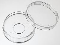
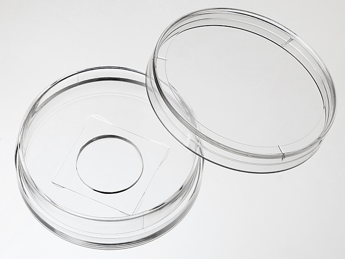
35 mm Glass bottom dish with 14 mm micro-well #1.5 cover glass


35mm glass bottom dish, dish size 35mm, well size 14mm, #1.5 cover glass(0.16mm-0.19mm). Designed for high resolution imaging such as confocal microscopy.
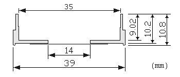
Note: We found that a small percentage of microscope adapters are too small for our 35 mm glass bottom dishes. Please check carefully the dimension diagram. If your adapter is too small, you should use our 29 mm glass bottom dish instead.
207 cases in stock
Features:
- Suitable for long term tissue culture
- Manufactured in a class 100,000 clean room
- Dish made from virgin polystyrene, tissue culture treated.
- German cover glass of superior optical quality
- A USP class VI adhesive is used to assemble the cover glass and the dish.
- Packed in easy to open peelable bag
- Sterilized by Gamma radiation.
Suitable for:
- Differential Interference Contrast (DIC)
- Widefield Fluorescence
- Confocal Microscopy
- Two-Photon and Multiphoton Microscopy
- Fluorescence Recovery After Photobleaching (FRAP)
- Förster Resonance Energy Transfer (FRET)
- Fluorescence Lifetime Imaging Microscopy (FLIM)
- Total Internal Reflection Fluorescence (TIRF)
- Super-Resolution Microscopy
Recommended for:
- Confocal Microscopy
Technical specifications
» View technical specification of different coverslips.
| Coverslip | #1.5 cover glass (0.16 - 0.19 mm) |
|---|---|
| Temperature Range | -20°C to 50°C |
| Lid diameter(outer) | 40 mm |
Dimension diagram (units in mm)

Cited Publications before 2019 (13)
-
Optical sensor revealed abnormal nuclease spatial activity on cancer cell membrane
Yongliang Wang, et al., Journal of Biophotonics, 29 November 2018
Quote: "First, 100 µg/ml BSA-biotin (Bovine serum albumin labeled by biotin, A8549, Sigma-Aldrich) and 5 µg/ml fibronectin were incubated on a glass-bottom petri dish (D35-14-1.5-N, In Vitro Scientific) for 30 min." -
Wide-area all-optical neurophysiology in acute brain slices
Samouil L Farhi, et al., BioRxiv, October 11, 2018
Quote: "After 24 hours, cells were split onto Matrigel (Fisher Scientific 356234) coated glass bottom plates (In Vitro Scientific D35-14-1.5-N) and imaged 24 hours later." -
Fast dynamic in vivo monitoring of Erk activity at single cell resolution in DREKA zebrafish
V Mayr, et al., Front Cell Dev Biol. 2018; 6: 111.
Quote: "A2576-25G, Sigma-Aldrich Chemie GmbH, Steinheim, Germany) in a glass bottom imaging dish (D35-14-1.5-NJ, Cellvis, Mountain View, CA, USA). Images and time-lapse movies were recorded on a Leica SP8 X WLL confocal microscope system" -
MCU interacts with Miro1 to modulate mitochondrial functions in neurons
Robert F. Niescier,, Journal of Neuroscience 23 April 2018,
Quote: "Glass bottom dishes (Cellvis, D35-14-1.5-N) were prepared 109 as follows for all microscopy experiments. First, dishes were immersed in 1 M HCl at 55°C 110 for at least 4 hours" -
A Fluorescence Fluctuation Spectroscopy Assay of Protein-Protein Interactions at Cell-Cell Contacts
V Dunsing, et al., Jove 2018
Quote: "35-mm glass bottom dishes CellVis D35-14-1.5-N" -
Two-photon photoactivated voltage imaging in tissue with an Archaerhodopsin-derived reporter
Miao-Ping Chien, et al., Arxiv, 2017
Quote: "Glass-bottom dishes covalently modified with fibronectin Glass-bottom dishes (In Vitro Scientific, D35-14-1.5-N) were covalently modified with fibronectin to facilitate subsequent photochemical crosslinking of cells to the dish" -
Simultaneous Real-Time Measurement of the β-Cell Membrane Potential and Ca2+ Influx to Assess the Role of Potassium Channels on β-Cell Function
NC Vierra, et al., Potassium Channels pp 73-84, 2017
Quote: "Poly-d-lysine coated 35 mm glass-bottom dishes (CellVis #D35–14-1.5-N)" -
Copy-number and gene dependency analysis reveals partial copy loss of wild-type SF3B1 as a novel cancer vulnerability
Brenton R Paolella, et al., eLife
Quote: "Cells were plated on 35 mm glass bottom dishes with #1.5 cover glass (D35-14–1.5 N, In Vitro Scientific)." -
ntegrins outside focal adhesions transmit tensions during stable cell adhesion
Y Wang, et al., Scientific Reports
Quote: "Glass bottom petridish (D35-14-1.5-N, In Vitro Scientific) was incubated with 1 mg/ml BSA-biotin" -
A role for activated Cdc42 in glioblastoma multiforme invasion
Hidehiro Okura, et al., Oncotarget
Quote: "Spheroids were then transferred to a 35 mm glass bottom imaging dish (In Vitro Scientific. Cat# D35-14-1.5-N) and embedded within BD Matrigel Basement Membrane Matrix"
View all publications citing "35 mm Glass bottom dish with 14 mm micro-well #1.5 cover glass".


