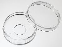
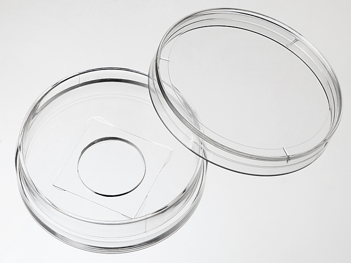
35 mm Glass bottom dish with 14 mm micro-well #1 cover glass


35 mm glass bottom dish, dish size 35mm, well size 14mm, #1 cover glass(0.13mm-0.16mm).
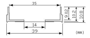
Note: We found that a small percentage of microscope adapters are too small for our 35 mm glass bottom dishes. Please check carefully the dimension diagram. If your adapter is too small, you should use our 29 mm glass bottom dish instead.
23 cases in stock
Features:
- Suitable for long term tissue culture
- Manufactured in a class 100,000 clean room
- Dish made from virgin polystyrene, tissue culture treated.
- German cover glass of superior optical quality
- A USP class VI adhesive is used to assemble the cover glass and the dish.
- Packed in easy to open peelable bag
- Sterilized by Gamma radiation.
Suitable for:
- Differential Interference Contrast (DIC)
- Widefield Fluorescence
- Confocal Microscopy
- Two-Photon and Multiphoton Microscopy
- Fluorescence Recovery After Photobleaching (FRAP)
- Förster Resonance Energy Transfer (FRET)
- Fluorescence Lifetime Imaging Microscopy (FLIM)
- Total Internal Reflection Fluorescence (TIRF)
- Super-Resolution Microscopy
Technical specifications
» View technical specification of different coverslips.
| Coverslip | #1 cover glass (0.13 - 0.16 mm) |
|---|---|
| Temperature Range | -20°C to 50°C |
| Lid diameter(outer) | 40 mm |
Dimension diagram (units in mm)

Cited Publications before 2019 (3)
-
Delivery of Fluorescent Probes Using Streptolysin O for Fluorescence Microscopy of Living Cells
KW Teng, et al., Current Protocols, Volume93, Issue1 August 2018
Quote: "Adherent cells are typically subcultured on a 35‐mm glass‐bottom dish with 14‐mm microwells (eg, Cellvis, cat. no. D35‐14‐1‐N) 2 days before the permeabilization experiment" -
Optimization of cerebellar purkinje neuron cultures and development of a plasmid-based method for purkinje neuron-specific, miRNA-mediated protein knockdown
CJ Alexander, et al., Methods in Cell Biology, September 2015
Quote: "Coat glass-bottom, 14-mm aperture, 35 mm culture dishes (In vitro Scientific D35-14-1-N) with 0.01% (w/v) poly-l-lysine (PLL) (Sigma P8920) supplemented with 1 mg/mL fibronectin" -
A Novel Method for Localizing Reporter Fluorescent Beads Near the Cell Culture Surface for Traction Force Microscopy
Samantha G. Knoll, et al., JoVE Bioengineering, 9/16/2014, Issue 91
Quote: "Materials. 35 mm Glass bottom dish with 14 mm micro-well #1 cover glass, In Vitro Scientific, D35-14-1-N"


