29 mm Glass bottom dish with 20 mm micro-well #0 cover glass
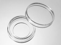

29 mm glass bottom dish, dish size 29mm, well size 20mm, #0 glass(0.085-0.115mm). Designed for high resolution imaging such as confocal microscopy.
10 cases in stock
Features:
- Suitable for long term tissue culture
- Manufactured in a class 100,000 clean room
- Dish made from virgin polystyrene, tissue culture treated.
- German cover glass of superior optical quality
- A USP class VI adhesive is used to assemble the cover glass and the dish.
- Packed in easy to open peelable bag
- Sterilized by Gamma radiation.
Suitable for:
- Differential Interference Contrast (DIC)
- Widefield Fluorescence
- Confocal Microscopy
- Two-Photon and Multiphoton Microscopy
- Fluorescence Recovery After Photobleaching (FRAP)
- Förster Resonance Energy Transfer (FRET)
- Fluorescence Lifetime Imaging Microscopy (FLIM)
- Total Internal Reflection Fluorescence (TIRF)
- Super-Resolution Microscopy
Technical specifications
» View technical specification of different coverslips.
| Coverslip | #0 cover glass (0.085-0.115 mm) |
|---|---|
| Temperature Range | -20°C to 50°C |
| Lid diameter (outer) | 34.3 mm |
Dimension diagram (units in mm)

Cited Publications before 2019 (5)
-
The effect of substrate bulk stiffness on focal and fibrillar adhesion formation in human abdominal aortic endothelial cells
H Hassanisaber, et al., Materials Science and Engineering: C Available online 29 December 2018
Quote: "Prior to imaging, the samples were washed three times with 1× PBS and mounted on 29 mm glass-bottom petri-dishes (Cellvis, D29-20-0-N, California, USA) on 100 μl of 50% (v/v) Glycerol (Thermo Fisher Scientific, G33) in 1× PBS" -
In Vivo Superresolution Imaging of Neuronal Structure in the Mouse Brain
BE Urban, et al., IEEE Transactions on Biomedical Engineering ( Volume: 65 , Issue: 1 , Jan. 2018 )
Quote: "Before the solution solidified, 1 mL of the mixture was placed in a 29 mm glass bottom dish with a 20 mm bottom well (D29-20-0-N, In Vitro Scientific) and immediately placed in a 4 °C environment to solidify" -
In vivo super-resolution imaging of neuronal structure in the mouse brain
BE Urban, et al., IEEE Transactions on Biomedic, Volume: 65 Issue: 1
Quote: "1 mL of the mixture was placed in a 29 mm glass bottom dish with a 20 mm bottom well (D29-20-0-N, In Vitro Scientific)" -
Spatiotemporal Stability of Neonatal Rat Cardiomyocyte Monolayers Spontaneous Activity Is Dependent on the Culture Substrate
J Boudreau-Béland, et al., Plos One, Jan 2, 2015
Quote: "Isolated cells (enriched cardiomyocytes) were counted and seeded at a density of 10 6 cells/mL in 20-mm diameter glass-bottom culture dishes (D29-20-0-N, In Vitro Scientific, Sunnyvale, CA, USA) pre-coated (on glass or on PDMS)" -
Multicellular automaticity of cardiac cell monolayers: effects of density and spatial distribution of pacemaker cells
JE Duverger, et al., New Journal of Physics, Volume 16, November 2014
Quote: "Isolated cells (enriched cardiomyocytes) were counted and seeded at a density of 1 × 10 6 cells mL −1 in 29 mm diameter glass-bottom culture dishes (D29-20-0-N, In vitro Scientific, Sunnyvale, CA) pre-coated with 0.2% gelatine and 0.001 25% fibronectin solution"