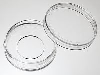
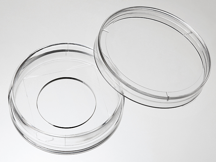
35 mm Glass bottom dish with 20 mm micro-well #1.5 cover glass


35mm glass bottom dish, dish size 35 mm, well size 20mm, #1.5 cover glass(0.16-0.19mm). Designed for high resolution imaging such as confocal microscopy.
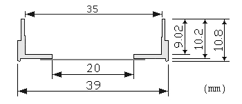
Note: We found that a small percentage of microscope adapters are too small for our 35 mm glass bottom dishes. Please check carefully the dimension diagram. If your adapter is too small, you should use our 29 mm glass bottom dish instead.
189 cases in stock
Features:
- Suitable for long term tissue culture
- Manufactured in a class 100,000 clean room
- Dish made from virgin polystyrene, tissue culture treated.
- German cover glass of superior optical quality
- A USP class VI adhesive is used to assemble the cover glass and the dish.
- Packed in easy to open peelable bag
- Sterilized by Gamma radiation.
Suitable for:
- Differential Interference Contrast (DIC)
- Widefield Fluorescence
- Confocal Microscopy
- Two-Photon and Multiphoton Microscopy
- Fluorescence Recovery After Photobleaching (FRAP)
- Förster Resonance Energy Transfer (FRET)
- Fluorescence Lifetime Imaging Microscopy (FLIM)
- Total Internal Reflection Fluorescence (TIRF)
- Super-Resolution Microscopy
Recommended for:
- Confocal Microscopy
Technical specifications
» View technical specification of different coverslips.
| Coverslip | #1.5 cover glass (0.16 - 0.19 mm) |
|---|---|
| Temperature Range | -20°C to 50°C |
| Lid diameter(outer) | 40 mm |
Dimension diagram (units in mm)

Cited Publications before 2019 (20)
-
A high-avidity biosensor reveals plasma membrane PI(3,4)P2 is predominantly a class I PI3K signaling product
Brady D. Goulden, et al., JCB, December 27, 2018
Quote: "For transfection, cells were seeded in 35-mm tissue culture dishes with 20-mm number 1.5 cover glass apertures (CellVis) coated with 5 µg fibronectin (Life Technologies 33016-015)" -
Mechanical loading of desmosomes depends on the magnitude and orientation of external stress
Andrew J. Price, et al., Nature Communications volume 9, Article number: 5284 (2018)
Quote: "For imaging experiments, MDCK cell lines were treated with 0.1 μg mL−1 (DPI-ctrl) or 0.5 μg mL−1 (DPI-TS and photometric controls) doxycycline in order to achieve similar levels of construct expression for the cell lines, and plated onto collagen-coated coverslips (Cellvis, D35-20-1.5-N) 48 hours (h) prior to imaging." -
Spatial Proliferation of Epithelial Cells Is Regulated by E-Cadherin Force
A Mohan, et al., Biophysical Jopurnal, Volume 115, Issue 5, 4 September 2018, Pages 853-864
Quote: "FRET imaging and analysis. Cells were seeded onto a 35 mm dish with a 1.5 NA (numerical aperture) 20-mm glass-bottom coverslip dishes (Cellvis, Sunnyvale, CA) that were precoated with 40 μg/mL fibronectin" -
Using Nesprin Tension Sensors to Measure Force on the LINC Complex
PT Arsenovic, et al., The LINC Complex pp 59-71
Quote: "For all imaging experiments, cells are grown on #1.5 coverglass bottom dishes (Cellvis D35-20-1.5-N)." -
Induction of oxidative stress by long-term treatment of live HEK293 cells with therapeutic concentration of lithium is associated with down-regulation of δ-opioid receptor amount and function
M Vosahlikova, et al., Biochemical Pharmacology Volume 154, August 2018, Pages 452-463
Quote: "The 35 mm glass bottom dish, dish size 35 mm, well size 20 mm, #1.5 cover glass (D35-20-1.5-N) was purchased from Cellvis (Mountain View, California, USA)" -
A cell surface display fluorescent biosensor for measuring MMP14 activity in real-time
lexander Braun, et al., Nature: Scientific Reportsvolume 8, Article number: 5916 (2018)
Quote: "For live imaging, HUVECs were cultured at 100,000 cells/dish on 10 µg/mL fibronectin coated 35 mm glass-bottom dishes (Cellvis, cat#: D35-20-1.5-N)" -
SAC1 degrades its lipid substrate PtdIns4P in the endoplasmic reticulum to maintain a steep chemical gradient with donor membranes
JP Zewe, et al., Elife, 2018
Quote: "For experiments, cells were seeded at 12.5-50% confluence on 10 μg/ml fibronectin (ThermoFisher 33016-015)-coated 35 mm dishes containing 20 mm #1.5 glass bottoms (CellVis D35-22-1.5-N) in 2 ml growth medium." -
Measuring mitotic forces
AY Anna, et al., Methods in Cell Biology Volume 144, 2018, Pages 165-184
Quote: "For semiadherent Drosophila S2 cells, the cells are seeded on a concanavalin A coated glass bottom 35 mm dish (Cellvis, Part No. D35-20-1.5-N) at ~ 70% confluency" -
Effect of therapeutic concentration of lithium on live HEK293 cells; increase of Na+/K+-ATPase, change of overall protein composition and alteration of surface layer of plasma membrane
Miroslava Vosahlikova, et al., Biochimica et Biophysica Acta
Quote: "35 mm glass bottom dishes, dish size 35 mm, well size 20 mm, #1.5 cover glass (D35-20-1.5-N) were purchased from Cellvis" -
A Protocol for Using Förster Resonance Energy Transfer (FRET)-force Biosensors to Measure Mechanical Forces across the Nuclear LINC Complex
PT Arsenovic, et al., Jove
Quote: "glass-bottom microscope viewing dish, In Vitro Scientific, D35-20-1.5-N, 35 mm Dish with 20 mm Bottom Well #1.5 glass."
View all publications citing "35 mm Glass bottom dish with 20 mm micro-well #1.5 cover glass".


